
Guideline 7.1 Routine blood loss
We suggest that during washback, dialysis lines and dialyser are observed to ensure residual blood loss is kept to a minimum. [2C]
Rationale
A small amount of blood loss occurs during normal haemodialysis, for example due to blood retained in the dialyser and circuit after washback, and bleeding into the dressing over needling sites, but there is no clear consensus as to what constitutes a ‘normal’ quantity of blood loss due to dialysis. The literature on minimising blood loss during haemodialysis is sparse, and much of the evidence is of limited quality.
The weighed gauze method has been to quantify bleeding after removal of needles, with ‘excessive’ defined as blood soaked gauze weighing over 4g [1]. Excessive bleeding has been associated with poor outcomes, for example in a study of 4152 dialysis sessions in 143 patients, Lin found that excessive bleeding following dialysis needle removal occurred regularly and was associated with lower haemoglobin levels [2,3]. Kalantar Zadeh suggested patients can lose up to 3g iron per year, with one gram being lost in the lines and dialyser, and a further gram lost in blood sampling [4]. Though it is unclear how they are derived, these estimates suggest that up to 20ml per session may be normal.
In a comparison of buttonhole versus rope ladder cannulation in 33 patients, Verhallen found no difference in bleeding times after needle removal between the two techniques [5]. Various suggestions have been made, for example, McCann suggested needling at an angle of 25 degrees [6], and Fruits suggested flushing the arterial dialysis needle with saline, and reducing the amount of blood drawn for testing, but none of these measures is well supported by clinical evidence [7]. Currently, there is insufficient evidence to support any recommendations regarding blood preservation and management of vascular access.
Clotting of the dialysis circuit leads to much greater blood loss than is routine. Adequate but safe anticoagulation is an important component of prevention, and is covered elsewhere in this guideline, but regular monitoring during dialysis and observation of the colour of the lines and dialyser post dialysis also play a role. This concept is supported in literature; for example, Kalocheritis noted the contribution of this type of blood loss to anaemia and the relevance of human factors [8]. Reasonable consensus, therefore, supports the importance of nursing observation, particularly during washback.
No evidence was found regarding the effects of excessive blood sampling on blood loss. Daugirdas and Tattersall point out that on line measurement of adequacy may reduce the need for blood sampling, but describe the benefits mainly in respect of cost and staff time [9]. However, ensuring that blood samples are taken only when required for routine monitoring or for additional diagnostic indications is perhaps obvious common sense.
References
- Awobusuyi JO, Mapayi FA, Adedolapo A. Blood loss during vascular access cannulation: Quantification using the weighed gauze and drape method. Haemodialysis International. 2008;12:90 93.
- Lin CL, Chen HY, Huang SC, Hsu SP, Pai MF, Peng YS, Chiu YL. Increased blood loss from access cannulation site during haemodialysis is associated with anemia and arteriovenous graft use. Therapeutic Apheresis and Dialysis. 2014;18(1):51 56.
- Tsai WC, Chen HY, Lin CL, Huang SC, Hsu SP, Pai MF, Peng YS, Chiu YL. Excessive cannulation site bleeding predicts long term all cause mortality in chronic haemodialysis patients. Therapeutic Apheresis and Dialysis. 2015;9(5):486–490.
- Kalantar Zadeh K, Streja E, Miller JE, Nissenson AR. Intravenous iron versus erythropoietin stimulating agents: Friends or foes in treating chronic kidney disease anemia. Adv Chronic Kidney Dis. 2009;16(2):143 51.
- Verhallen AM, Kooistra MP, van Jaarsveld BC. Cannulating in haemodialysis: rope ladder or buttonhole technique? Nephrol Dial Transplant. 2007;22:2601–2604.
- McCann M, Einarsdóttir H, Van Waeleghem JP, Murphy F, Sedgewick J. Cannulation practice patterns and outcomes in vascular access. J Ren Care. 2008;34(2):77 84.
- Fruits RG, Kinney R, Hegeman T. Maintaining iron balance by decreasing blood loss. ANNA Journal. 1992;19(2):156.
- Kalocheretis P, Vlamis I, Belesi C, Makriniotou I, Zerbala S, Savidou E, Zorbas S, Arvanitis N, Iatrou C. Residual blood loss in single use dialyzers: effect of different membranes and flux. Int J Artif Organs. 2006;29(3):286 92.
- Daugirdas JT, Tattersall JE. Automated monitoring of haemodialysis adequacy by dialysis machines: potential benefits to patients and cost savings. Kidney International. 2010;78:833 835.
Guideline 7.2 Disconnection haemorrhage
We recommend maintaining awareness of the risk of disconnection, the limitations of pressure alarms, and the importance of direct observation through a program of education, including patients and carers. [1D]
We suggest regular assessment of individual risk, so that high risk patients can have enhanced monitoring, which could include specific devices. [2B]
Rationale
Disconnection leading to haemorrhage may occur at any part of the dialysis circuit, though venous needle dislodgement may be the most frequent and serious, with rapid blood loss occurring at the rate of the blood flow pump until it is detected. Disconnection incidents are thought to be uncommon, but the true prevalence is uncertain due to inconsistent reporting. Once detected, management begins with haemostasis and fluid resuscitation, as with any major haemorrhage. The literature focuses primarily on methods to minimise risk and enhance detection, with publications available from the EDTNA/ERCA and the American Nephrology Nurses Association.
Variability in human processes is recognised as an important factor, and most units have established protocols to ensure consistency in aspects of care, such as taping needles in position to minimise the chance of disconnection.
Dialysis machines have several types of safety monitors, and if disconnection does occur, the drop in pressure should be detected and cause the machine to alarm. However, it has been repeatedly demonstrated that these alarms cannot be relied on to detect all cases. The use of asymmetric windows (such as 30 to +70 mmHg) may help maximise the detection of disconnection while minimising alarms from increases in pressure at the venous needle.
Because machine alarms cannot be relied upon entirely, direct observation remains crucial, involving vigilance on the part of nursing staff and unit management, so that lines of sight are not obscured, patients are not dialysing alone, and their vascular access sites are not covered. Given the low prevalence of disconnection, complacency may develop; therefore, continuous education is advocated to ensure awareness among healthcare staff, patients, and their carers.
Risk of disconnection is greater in some patients, and enhanced monitoring may be appropriate based on individual risk assessment. Simply placing patients closer to the nursing desk may be sufficient, but reliable monitoring can also be achieved by using blood loss detection devices, which typically are secured at the vascular access site and alarm on the detection of blood. Device monitoring may be appropriate for high risk patients, such as those who are confused or agitated, and may play a larger role in home haemodialysis programs. One interventional study showed that the use of blood loss detection devices increased the self reported feeling of safety among nursing staff.
References
- Van Waeleghem JP, Chamney M, Lindley EJ, Pancírová J. Venous needle dislodgement: how to minimise the risks. J Ren Care. 2008 Dec;34(4):163 8. doi: 10.1111/j.1755 6686.2008.00047.x.
- Axley B, Speranza Reid J, Williams H. Venous needle dislodgement in patients on haemodialysis. Nephrol Nurs J. 2012 Nov Dec;39(6):435 45; quiz 446.
- Zeigler SA. Prevent dangerous haemodialysis catheter disconnections. Nursing. 2007 Mar;37(3):70.
- International Standard IEC 60601 2 16. Medical electrical equipment part 2 16: Particular requirements for the safety of haemodialysis, haemodiafiltration and haemofiltration equipment. 2008. (available at: https://webstore.iec.ch/publication/2619)
- Allcock K, Jagannathan B, Hood CJ, Marshall MR. Exsanguination of a home haemodialysis patient as a result of misconnected blood lines during the wash back procedure: a case report. BMC Nephrol. 2012 May 15;13:28. doi: 10.1186/1471 2369 13 28.
- Ribitsch W, Schilcher G, Hafner Giessauf H, Krisper P, Horina JH, Rosenkranz AR, Schneditz D. Prevalence of detectable venous pressure drops expected with venous needle dislodgement. Semin Dial. 2014 Sep Oct;27(5):507 11. doi: 10.1111/sdi.12169.
- Polaschegg HD. Venous needle dislodgement: the pitfalls of venous pressure measurement and possible alternatives, a review. J Ren Care. 2010 Mar;36(1):41 8. doi: 10.1111/j.1755 6686.2010.00142.x.
- Garrick R, Morey R. Dialysis Facility Safety: Processes and Opportunities. Semin Dial. 2015 Sep Oct;28(5):514 24. doi: 10.1111/sdi.12395.
- Takeuchi A, Miwa T, Shirataka M, Sawada M, Imaizumi H, Sugibuchi H, Ikeda N. Non cladding optical fiber is available for detecting blood or liquids. J Clin Monit Comput. 2010 Oct;24(5):363 70. doi: 10.1007/s10877 010 9255 z.
- NHS PASA Centre for Evidence based Purchasing. Evidence Review: Redsense blood loss detection device for venous needle dislodgement monitoring in haemodialysis. 2009. (available at: http://www.renal.org/docs/default source/patient safety docs/patient safety reports/redsense cep08050 mar 09.pdf?sfvrsn=2)
- Pauly RP, Eastwood DO, Marshall MR. Patient safety in home haemodialysis: quality assurance and serious adverse events in the home setting. Hemodial Int. 2015 Apr;19 Suppl 1:S59 70. doi: 10.1111/hdi.12248.
- Wong B, Zimmerman D, Reintjes F, Courtney M, Klarenbach S, Dowling G, Pauly RP. Procedure related serious adverse events among home haemodialysis patients: a quality assurance perspective. Am J Kidney Dis. 2014 Feb;63(2):251 8. doi: 10.1053/j.ajkd.2013.07.009.
- Hawley CM, Jeffries J, Nearhos J, Van Eps C. Complications of home haemodialysis. Hemodial Int. 2008 Jul;12 Suppl 1:S21 5. doi: 10.1111/j.1542 4758.2008.00291.x.
- Tennankore KK, d’Gama C, Faratro R, Fung S, Wong E, Chan CT. Adverse technical events in home haemodialysis. Am J Kidney Dis. 2015 Jan;65(1):116 21. doi: 10.1053/j.ajkd.2014.08.013.
- Ahlmén J, Gydell KH, Hadimeri H, Hernandez I, Rogland B, Strömbom U. A new safety device for haemodialysis. Hemodial Int. 2008 Apr;12(2):264 7. doi: 10.1111/j.1542 4758.2008.00263.x.
Guideline 7.3 Immune reactions during dialysis
We recommend that dialysis staff should be aware of the features and management of dialysis reactions, and should have access to a range of dialyser types. [1C]
Rationale
From the early 1980s, reports described abrupt clinical reactions occurring soon after the onset of dialysis. These have traditionally been classified into two types:
Type A reactions were reported to affect less than 1% of patients per year, often recurring in the same patient, with onset within the first few minutes of dialysis. Primarily occurring with first use (rather than re used dialysers), the features were quite ‘anaphylactic’ in nature (itching, flushing, bronchospasm, hypotension, sometimes with burning at the access site) and occasionally severe, with cardiac arrest described. These reactions were associated with eosinophilia and were mainly caused by residual ethylene dioxide used to sterilize membranes, with antibodies detectable in some cases. Similar reactions were seen with polyacrylonitrile membranes, especially in ACE inhibitor treated patients (due to increased kinin activation) and with hydrogen peroxide treated re used membranes. Immediate cessation of dialysis was typically necessary, along with anaphylaxis type treatment. Extra rinsing or a change of membrane sterilization often prevented reoccurrence.
Type B reactions, reportedly more common, occurred later in the dialysis session and were generally less severe, improving with continued dialysis. Characterized mainly by chest and back pain (also occasionally vomiting, breathlessness, and hypotension), these reactions were caused by complement activation and pulmonary cell sequestration, associated with transient reductions in circulating white cells, and were linked to the ‘bio incompatibility’ of cellulose based membranes.
Dialyser re use, ethylene dioxide sterilization, and unmodified cellulose membranes are now uncommon, and as dialysis practices have evolved, the epidemiology of these reactions has changed. In modern practice, dialysis reactions are uncommon but still occur, including polysulfone allergy, heparin allergy, and isolated thrombocytopenia.
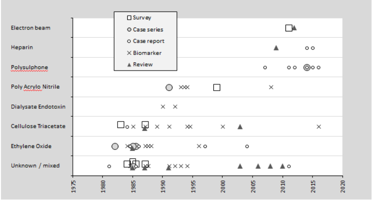
Figure 2. Literature timeline showing the changing epidemiology of dialysis reactions.
Reactions with ‘Type A’ (anaphylactic) features continue with polysulfone membranes, though variants include those with fever as the predominant symptom. Eosinophilia is an important indicator, though not always present, and other blood tests (tryptase, total IgE) may be useful. The diagnostic hallmark is resolution of the syndrome after changing the membrane type. Anaphylaxis treatments are often given, and steroid pre treatment is sometimes used before dialysis sessions. Stopping ACE inhibitors may also reduce severity.
Reactions to intra dialytic heparin range from asymptomatic to a serotonin like syndrome with breathlessness, flushing, and often hypertension. These are usually but not always associated with thrombocytopenia (persisting between dialysis sessions), and thrombotic complications may occur. Transient asymptomatic thrombocytopenia has also been reported, often recovering between dialysis sessions. This reaction is associated with electron beam membrane sterilization, though the mechanism is unknown.
Several complications other than dialyser reactions may present with similar symptoms, including bacteraemia and hypovolaemia. Rare complications include disequilibrium, air embolism, and chloramine or hard water syndromes, with water purification complications potentially more common in home haemodialysis settings.
References
- Nicholls AJ, Platts MM. Anaphylactoid reactions due to haemodialysis, haemofiltration, or membrane plasma separation. Br Med J (Clin Res Ed). 1982 Dec 4;285(6355):1607 9.
- Nicholls AJ. Hypersensitivity to haemodialysis: the United Kingdom experience. Artif Organs. 1987 Apr;11(2):87 9.
- Grammer LC, Roberts M, Nicholls AJ, Platts MM, Patterson R. IgE against ethylene oxide altered human serum albumin in patients who have had acute dialysis reactions. J Allergy Clin Immunol. 1984 Oct;74(4 Pt 1):544 6.
- Schaefer RM, Fink E, Schaefer L, Barkhausen R, Kulzer P, Heidland A. Role of bradykinin in anaphylactoid reactions during haemodialysis with AN69 dialyzers. Am J Nephrol. 1993;13(6):473 7.
- Amore A, Guarnieri G, Atti M, Schena FP, Coppo R. Use of alkaline rinsing solution to prevent hypersensitivity reactions during haemodialysis: data from a multicentre retrospective analysis. J Nephrol. 1999 Nov Dec;12(6):383 9.
- Sánchez Villanueva RJ, González E, Quirce S, Díaz R, Alvarez L, Menéndez D, Rodríguez Gayo L, Bajo MA, Selgas R. Hypersensitivity reactions to synthetic haemodialysis membranes. Nefrologia. 2014;34(4):520 5.
- Hildebrand S, Corbett R, Duncan N, Ashby D. Increased prevalence of eosinophilia in a haemodialysis population: Longitudinal and case control studies. Hemodial Int. 2016 Jul;20(3):414 20.
- Kiaii M, Djurdjev O, Farah M, Levin A, Jung B, MacRae J. Use of electron beam sterilized haemodialysis membranes and risk of thrombocytopenia. JAMA. 2011 Oct 19;306(15):1679 87.